
Page 117 - Ship Construction.DJ Eyres 6Ed
P. 117
Ch10-H8070.fm Page 106 Wednesday, October 18, 2006 7:37 AM
106 Ship Construction
the particles will concentrate at this point where there is an alteration in the
magnetic field. A dye penetrant will also show up a surface flaw if it remains
after the casting has been washed following the application of the dye. To
aid the detection of a surface crack the dye penetrant used is often lumi-
nous and is revealed under an ultra-violet light.
Visual inspection of welds is routine procedure, and surface defects are
soon noticed by the experienced inspector and surveyor. Incorrect bead
--- ใช้เพื่อการศึกษาเท่านั้น---
shape, high spatter, undercutting, bad stop and start points, incorrect align-
ment, and surface cracks are all faults which may be observed at the surface.
Sub-surface and internal defects are not observed, but the cost of visual
งานห้องสมุด ศูนย์ฝกพาณิชย์นาวี
inspection is low, and it can be very effective where examination is made
before, during, and after welding.
The principle of radiographic inspection is simply to subject a material to
radiation from one side, and record the radiation emitted from the opposite
side. Any obstacle in the path of the radiation will affect the radiation density
emitted and may be recorded. As radiation will expose photographic plate,
for all practical weld test purposes this is used to record the consistency of the
weld metal. The photographic plate records changes in radiation density emit-
ted; for example a void will show up as a darker shadow on the radiograph.
Either X-ray or gamma ray devices may be used to provide the source of
ึ
radiation. X-ray equipment consists of a high voltage power source (50 to
400kV) which is used to provide potential between a cathode and target
anode in a glass vacuum tube. Only a small percentage of this energy is con-
verted to X-rays, so that large amounts of heat have to be conducted away
from the target. From the target the X-rays are projected out of the tube
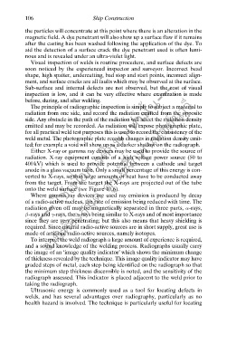
onto the weld surface (see Figure 10.6).
Where gamma ray devices are used ray emission is produced by decay
of a radio-active nucleus, the rate of emission being reduced with time. The
radiation given off may be magnetically separated in three parts, a-rays,
b-rays and g-rays, the g-rays being similar to X-rays and of most importance
since they are very penetrating; but this also means that heavy shielding is
required. Since natural radio-active sources are in short supply, great use is
made of artificial radio-active sources, namely isotopes.
To interpret the weld radiograph a large amount of experience is required,
and a sound knowledge of the welding process. Radiographs usually carry
the image of an ‘image quality indicator’ which shows the minimum change
of thickness revealed by the technique. This image quality indicator may have
graded steps of metal, each step being identified on the radiograph so that
the minimum step thickness discernible is noted, and the sensitivity of the
radiograph assessed. This indicator is placed adjacent to the weld prior to
taking the radiograph.
Ultrasonic energy is commonly used as a tool for locating defects in
welds, and has several advantages over radiography, particularly as no
health hazard is involved. The technique is particularly useful for locating